Diagram Of Upper Leg Muscles And Tendons - Inner Thigh Muscles Anatomy Anatomy Drawing Diagram - In other words, this page excludes information about the calf.
Diagram Of Upper Leg Muscles And Tendons - Inner Thigh Muscles Anatomy Anatomy Drawing Diagram - In other words, this page excludes information about the calf.. The fibers converge into a tendon which passes under the foot and attaches to the medial side of the foot. Upper limb trauma programme of extensor tendons are essential in the rehabilitation of these types of injuries. The muscular system consists of the skeletal muscles and their associated structures. Tendonitis is usually seen after excessive repetitive movement with which the tendon gradually becomes tighter until the fibers start to tear. Plantarflexes the foot at the ankle joint.
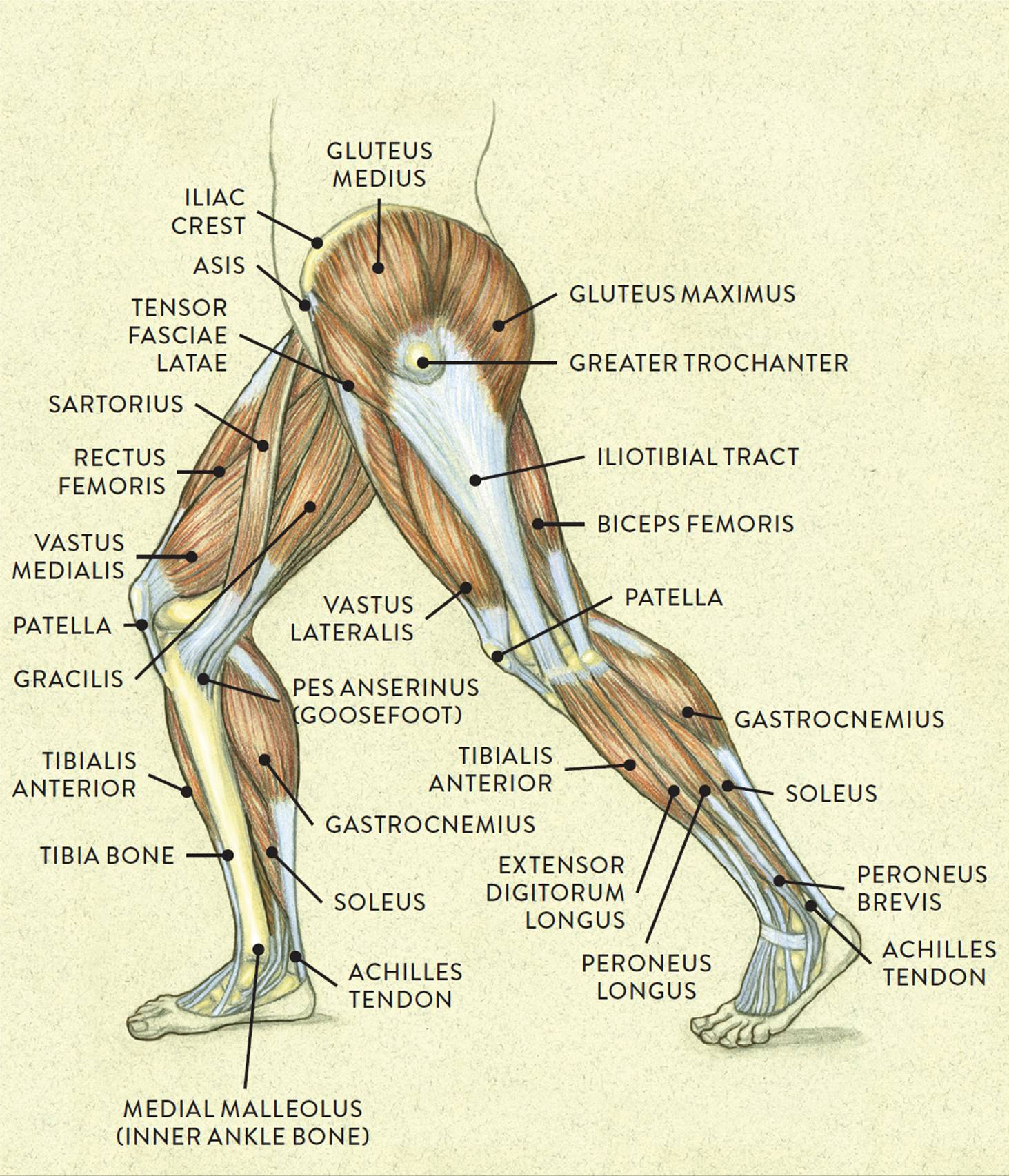
The core muscles are those in the abdomen, back, and pelvis, and they also stabilize the body and assist in tasks, such as lifting weights. Specifically, this page discusses all the major muscle groups of the upper leg. Originates from the fibula and tibia. Leg muscles are another story. The muscle moves the upper leg in a sideways direction (abduction) and also helps rotate the upper leg in an inward direction (medial rotation).
Concept 3d Illustration Front Upper Leg Human Anatomy Stock Illustration Illustration Of Anatomy Gastrocnemius 99931201 from thumbs.dreamstime.com Human muscle system, the muscles of the human body that work the skeletal system, that are under voluntary control, and broadly considered, human muscle—like the muscles of all vertebrates—is often divided into striated muscle, smooth skeletal muscles are attached to the bones by tendons. Leg muscles are another story. Sciatic nerve present gluteus medius abducts & rotates thigh medially gluteus minimus deep to medius; A tendon is the end part of a muscle that attaches the muscle to the bone. Tendonitis is usually seen after excessive repetitive movement with which the tendon gradually becomes tighter until the fibers start to tear. But there's a wide range of sizes and muscle makeup among people that even experts debate. Muscles of the leg include muscles of the thigh and foot. Tendons are cords made of tough tissue, and they work as special connector pieces between bone and muscle.
Other areas where tendonitis occurs include the hips and ankles.
One more example is the large muscle group of the quadriceps, located on the front of the upper leg. Extends & rotates thigh laterally; Originates from the fibula and tibia. Anterior, lateral and posterior compartment. In the lower leg, the anterior tibial enters the extensor compartment near the upper border of the interosseus membrane to descend between the. Tendonitis is usually seen after excessive repetitive movement with which the tendon gradually becomes tighter until the fibers start to tear. Leg muscles are another story. Muscle tendons stretch over joints and contribute to joint stability. Both peroneal tendons then course anteriorly toward the peroneal trochlea of the lateral calcaneum, at which point the longus tendon runs inferiorly to the peroneal. Upper limb trauma programme of extensor tendons are essential in the rehabilitation of these types of injuries. Sartorius muscle appears from the anterior superior iliac spine and upper half of the notch immediately below it. But there's a wide range of sizes and muscle makeup among people that even experts debate. Section editor dean taylor, md.
Each of these muscles is a discrete organ constructed of skeletal muscle tissue, blood vessels, tendons, and nerves. Related online courses on physioplus. Muscle tendons stretch over joints and contribute to joint stability. Anterior, lateral and posterior compartment. The leg muscles are organized in 3 groups:
Thigh Muscle Diagram Leg Muscles Diagram Muscle Diagram Leg Muscles from i.pinimg.com Tendons are cords made of tough tissue, and they work as special connector pieces between bone and muscle. Muscle tendons stretch over joints and contribute to joint stability. They depend greatly on our genes and what we do with them. But there's a wide range of sizes and muscle makeup among people that even experts debate. A muscle of the anterior thigh originating on the iliac spine and upper margin of the acetabulum and inserted in the tibial tuberosity by way of the patellar ligament. Human muscle system, the muscles of the human body that work the skeletal system, that are under voluntary control, and broadly considered, human muscle—like the muscles of all vertebrates—is often divided into striated muscle, smooth skeletal muscles are attached to the bones by tendons. A muscle along the outside of the leg that bends the foot out at the ankle. Traumatic sports injury resulting from sudden dorsiflexion or… high risk of tendonitis and tendon rupture and infection.
Abducts & rotates thigh medially.
The core muscles are those in the abdomen, back, and pelvis, and they also stabilize the body and assist in tasks, such as lifting weights. The peroneus longus muscle (also known as fibularis longus muscle) is one of the muscles of the lateral compartment of the leg. Plantarflexes the foot at the ankle joint. Many of the leg's muscles are also adapted to bipedalism, most substantially the gluteal muscles, the extensors of the knee joint, and the calf muscles.8. The muscles of the foot mainly customize and improve the actions of the long tendons and help fine movements of the toes. A muscle along the outside of the leg that bends the foot out at the ankle. Muscle tendons stretch over joints and contribute to joint stability. Other areas where tendonitis occurs include the hips and ankles. Lesson on the anatomy of the forearm: Muscles of the leg include muscles of the thigh and foot. Specifically, this page discusses all the major muscle groups of the upper leg. Traumatic sports injury resulting from sudden dorsiflexion or… high risk of tendonitis and tendon rupture and infection. A tendon is the end part of a muscle that attaches the muscle to the bone.
The fibers converge into a tendon which passes under the foot and attaches to the medial side of the foot. When muscles get tight, they are actually getting less pliable, meaning that they cannot lengthen properly and therefore restrict the motion of the joint they are connected to. Sartorius muscle appears from the anterior superior iliac spine and upper half of the notch immediately below it. One more example is the large muscle group of the quadriceps, located on the front of the upper leg. Muscles of the leg include muscles of the thigh and foot.
Muscles Of The Leg And Foot Classic Human Anatomy In Motion The Artist S Guide To The Dynamics Of Figure Drawing from doctorlib.info Other areas where tendonitis occurs include the hips and ankles. A tendon is the end part of a muscle that attaches the muscle to the bone. Related online courses on physioplus. Each of these muscles is a discrete organ constructed of skeletal muscle tissue, blood vessels, tendons, and nerves. Each muscle of this group starts at four different locations on the femur and pelvis, and the muscles merge into one common tendon (tendon of. This is where the gto comes into play. Specifically, this page discusses all the major muscle groups of the upper leg. Abducts & rotates thigh medially.
The biomechanical effects of stretching.
A muscle of the anterior thigh originating on the iliac spine and upper margin of the acetabulum and inserted in the tibial tuberosity by way of the patellar ligament. When muscles get tight, they are actually getting less pliable, meaning that they cannot lengthen properly and therefore restrict the motion of the joint they are connected to. The muscle ends in tendons and the tendons plug the muscle into bones. Sartorius muscle appears from the anterior superior iliac spine and upper half of the notch immediately below it. 3 s and s muscles of the upper leg brachioradialis radius (styloid process) gluteal group gluteus maximus largest; Muscles of the leg include muscles of the thigh and foot. This is where the gto comes into play. One more example is the large muscle group of the quadriceps, located on the front of the upper leg. Lesson on the anatomy of the forearm: At the lower leg, peroneus longus muscle injuries (e.g., denervation) along with retromalleolar tendon instability/subluxation will be discussed. The fibers converge into a tendon which passes under the foot and attaches to the medial side of the foot. By striking in at a 90 degree angle into the bone, pain and dysfunction will. The muscles of the foot mainly customize and improve the actions of the long tendons and help fine movements of the toes.
Tendons are cords made of tough tissue, and they work as special connector pieces between bone and muscle upper leg muscles and tendons. When muscles get tight, they are actually getting less pliable, meaning that they cannot lengthen properly and therefore restrict the motion of the joint they are connected to.


0 Komentar